


توضیحات
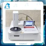
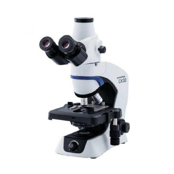
دستگاه میکروسکوپ متالوژی عبوری و انعکاسی مدل BX53M کمپانی Olympus ژاپن
میکروسکوپ متالورژی Olympus BX53M یک دستگاه حرفهای و پیشرفته از نمایندگی Olympus ژاپن است که به طور خاص برای آنالیزهای متالورژیکی طراحی شده است.
میکروسکوپ BX53M یک میکروسکوپ نوری برای مشاهده و آنالیز نمونههای فلزی و مواد غیرشفاف دیگر است. طراحی Modular (پیمانهای) آن امکان سفارشیسازی و تطبیق با نیازهای خاص کاربر را فراهم میکند. این مدل به دلیل وضوح تصویر فوق العاده، پایداری و ارگونومی عالی، یک انتخاب استاندارد در آزمایشگاههای کنترل کیفیت، تحقیق و توسعه و مراکز آموزشی است.
جهت کسب اطلاعات و مشخصات فنی میکروسکوپ متالوژی عبوری و انعکاسی مدل BX53M کاتالوگ زیر را مطالعه فرمایید.
ویژگیها میکروسکوپ متالوژی عبوری و انعکاسی مدل BX53M
۱. سیستم نوری UIS2 (Universal Infinity System)
-
این پیشرفتهترین سیستم نوری Olympus است که از اپتیک بینهایت (Infinity-corrected) استفاده میکند.
-
مزیت: وضوح (Resolution) و کنتراست بسیار بالا در تمامی بزرگنماییها
-
مزیت: ایجاد تصاویر مسطح (Flat Field) در سراسر میدان دید
-
مزیت: انعطافپذیری برای اضافه کردن انواع کنتراست و دتکتور بدون تاثیر بر کیفیت تصویر
۲. روشهای مشاهده (Observation Methods) جامع
این میکروسکوپ از تمامی تکنیکهای متالورژیکی ضروری پشتیبانی میکند:
-
reflected Light انعکاسی اصلیترین روش برای مشاهده نمونههای فلزی و غیرشفاف
-
Brightfield (BF): روش استاندارد برای مشاهده کلی ساختار
-
Darkfield (DF): برای مشاهده ترکها، حفرهها و مرزدانهها با کنتراست بالا
-
Polarized Light (POL): برای شناسایی فازهای غیرایزوتروپیک، اکسیدها و inclusions
-
Differential Interference Contrast (DIC): برای ایجاد کنتراست سهبعدی و مشاهده جزئیات ظریف سطح و مرز دانهها
-
Light (عبوری): این قابلیت به صورت اختیاری و برای نمونههای نیمه شفاف (مثلاً در زمینشناسی یا سرامیک) افزوده میشود.
۳. طراحی ارگونومیک و ضد خستگی (Ergonomics)
-
Focus Drive: موقعیت کانونی کردن در ارتفاعی مناسب قرار دارد و بسیار نرم است.
-
Stage Drive: میز نمونه (stage) بزرگ و ثابت است و نمونه به سمت چپ و راست حرکت میکند که برای کاربر راستدست یا چپدست قابل تنظیم است. این طراحی از برخورد کاربر با ابزارها جلوگیری میکند.
-
HD DIC: کنتراست تفاضلی با وضوح بالا برای کمکردن خستگی چشم
۴. سازگاری با دوربینهای دیجیتال و نرمافزار آنالیز تصویر
-
این میکروسکوپ به راحتی با دوربینهای دیجیتال Olympus (مانند سری UC90) و دیگر برندها سازگار است.
-
امکان اتصال به کامپیوتر و نرمافزارهای پیشرفته آنالیز تصویر برای موارد زیر وجود دارد:
-
اندازهگیری ابعادی (ذرات، لایهها و…)
-
آنالیز اندازه دانه (Grain Size) مطابق با استانداردهای ASTM E112 و…
-
تعیین درصد فازها (Phase Percentage)
-
ایجاد پانوراما (تصویربرداری از کل نمونه)
-
ایجاد گزارشهای خودکار
-
۵. منبع نور (Illumination)
-
معمولاً از یک منبع نور LED استفاده میکند.
-
مزایای نور LED: طول عمر بسیار بالا، پایداری نوری، مصرف انرژی پایین، تولید حرارت کم (که برای نمونهها مفید است) و روشنایی یکنواخت
۶. قابلیتهای پیشرفته (اختیاری)
-
Motorization: امکان موتورایز کردن فوکوس، چرخۀ کنتراست و stage برای انجام اتوماتیک tasks
-
DSM (Digital Slide Maker): برای ایجاد اسلایدهای دیجیتال از کل نمونه
کاربردهای اصلی میکروسکوپ متالوژی عبوری و انعکاسی مدل BX53M
از این میکروسکوپ در صنایع و زمینههای زیر به طور گسترده استفاده میشود:
-
متالورژی و علم مواد
-
بررسی ریزساختار فلزات و آلیاژها (فولاد، چدن، آلومینیوم، تیتانیوم و…)
-
تعیین اندازه دانه (Grain Size)
-
شناسایی و توزیع فازها (مانند کاربیدها در فولاد)
-
بررسی inclusions و ناخالصیها
-
ارزیابی پوششهای سطحی و عمق لایه سختشده
-
آنالیز شکست (Fractography)
-
-
صنعت خودروسازی و هوافضا
-
کنترل کیفیت قطعات تولیدی
-
آنالیز علل شکست قطعات
-
-
صنایع الکترونیک
-
بررسی اتصالات، bonding wires
-
قطعات نیمههادی
-
-
زمینشناسی
-
شناسایی کانیها و سنگها (با استفاده از روشهای پلاریزه و عبوری اختیاری)
-
-
سرامیک و کامپوزیتها
-
بررسی تخلخل، ترک و توزیع ذرات
-
-
تحقیقات آکادمیک
در دانشگاهها و مراکز پژوهشی برای مطالعه پیشرفته مواد
جمع بندی نهایی
میکروسکوپ Olympus BX53M را میتوان یک ابزار صنعتی تمامعیار و حرفهای دانست.
نقاط قوت:
-
کیفیت اپتیکال بینظیر: سیستم نوری UIS2 تصاویری با وضوح و کنتراست استثنایی ارائه میدهد.
-
انعطافپذیری و ماژولار بودن: میتوان آن را برای طیف وسیعی از کاربردهاکرد.
-
ساختار مستحکم و پایدار: برای محیطهای صنعتی و استفاده سنگین طراحی شده است.
-
کاربری آسان و ارگونومیک: امکان کار طولانیمدت بدون خستگی کاربر.
-
پشتیبانی قوی نرمافزاری و سختافزاری: قابلیت ارتقا به سیستمهای تمام اتوماتیک و آنالیز کمی تصویر.
” جهت استعلام قیمت میکروسکوپ متالوژی عبوری و انعکاسی مدل BX53M با مشاورین واحد فروش شرکت آراتجهیز فارمد ۸۸۴۰۱۷۰۰-۰۲۱ در ارتباط باشید. “
BX53M reflected light/Upright field Specification
| Optical system | UIS2 optical system (infinity-corrected) | ||
| Microscope frame | Illumination | Reflected/transmitted | |
| Focus | Stroke: 25 mm Fine stroke per rotation: 100 um Minimum graduation: 1 um With upper limit stopper, torque adjustment for coarse handle | ||
| Max. specimen height | 35 mm (w/o spacer) 75 mm (with BX3M-ARMAD) | ||
| Observation tube | Wide-field FN 22 | Inverted: binocular, trinocular, tilting binocular Erect: trinocular, tilting binocular | |
| Super-wide-field FN 26.5 | Inverted: trinocular Erect: trinocular, tilting trinocular | ||
| Reflected light illumination | Traditional observation technique | BX3M-RLAS-S Coded, white LED, BF/DF/DIC/POL/MIX FS, AS (with centering mechanism) BX3M-KMA-S White LED, BF/DIC/POL/MIX FS, AS (with centering mechanism) BX3M-RLA-S 100W/50W halogen lamp, white LED, BF/DF/DIC/POL/MIX/ FS, AS (with centering mechanism), BF/DF interlocking, ND filter | |
| – | |||
| Fluorescence | BX3M-URAS-S Coded, 100W mercury lamp, 4 position mirror unit turret, (standard: WB, WG, WU+BF etc) With FS, AS (with centering mechanism), with shutter mechanism | ||
| Transmitted light | White LED Abbe/long working distance condensers | ||
| Revolving nosepiece | For BF | Sextuple, centering sextuple, septuple, coded quintuple (optional motorized revolving nosepieces) | |
| For BF/DF | Sextuple, quintuple, centering quintuple, coded quintuple (optional motorized revolving nosepieces) | ||
| Stage | Coaxial left (right) handle stage: 7x52mm, with torque adjustment Large-size coaxial left (right) handle stage: 105x100mm, with locking mechanism in Y-axis Large-size coaxial right handle stage: 150x100mm, with torque adjustment and locking mechanism in Y-axis | ||
| Weight | Approx. 18.3 kg (Microscope frame 7.6 kg) | ||
Optical system: infinity corrected
Tripod: UPright /reflected light
Focus: travel: 25 mm, 100 µm per revolution, smallest division: 1 µm, with upper stop and torque adjustment for coarse adjustment
Max. height of object: 65 mm (without spacer), 105 mm (with spacer BX3M-ARMAD)
Observation tube: Wide field of view (F.N,22), inverted: Binocular with camera tube
Lighting: White light LED
Objective turret for bright field: 5-fold
Eyepiece: 10x/22 diopter adjustment on one side
Table (X x Y) 76 mm × 52 mm Coaxial table with left-hand drive with torque adjustment
Weight: approx. 15.8 kg (microscope stand 7.4 kg)
Modules included in delivery:
Tripods &mounts BX53MRF-S-1-3
Trinocular tube U-TR30-2-2
Eyepiece WHN10X-1-8
Eyepiece WHN10X-H-1-8
U-KMASreflected light condenser
Light source BX3M-LEDR
Objective turret U-5RE-2
Cross table U-SVRM Right-hand drive
Table insert plate U-MSSP
Connection cable UYCP
Objectives can be ordered separately; they are not included in the scope of delivery!
Please select your preferred accessories for this model: MPLFLN lenses
Modern microscopy made easy
With its modular concept, the BX3M series offers versatility for a wide range of applications in industry and materials research. Thanks to improved integration with OLYMPUS Stream software, BX53M microscopes enable smooth workflows for standard microscopy applications and digital imaging, from examination to reporting.
Easily use familiar procedures:
Simple light source
The light source minimises complicated steps that are usually required when working with a microscope. The adjustment knob on the front of the condenser allows the microscopy method to be selected conveniently. Users can quickly switch between the most commonly used methods of reflected light microscopy, from bright field to dark field or polarised light microscopy, to easily perform different types of analysis. In addition, simple polarised light microscopy can be adjusted by rotating the analyser.
Intuitive microscope controls:
Simple adjustment of the illumination field diaphragm and aperture diaphragm
Correct adjustment of the illumination field and aperture diaphragm ensures good image contrast and full utilisation of the numerical aperture of the objective. The Legend guides the user through the correct settings for the respective microscopy method and the objective used.
Fast focus adjustment
The focus scale on the tripod facilitates quick focusing. Users can roughly adjust the focus without looking through an eyepiece, saving time when examining objects of different heights.
Easy and ergonomic operation
Ergonomics is of paramount importance for all users. Whether a user is using their microscope on its own or in conjunction with OLYMPS Stream image analysis software, they will benefit from the ergonomic hand switches that clearly indicate the position of the hardware. The ergonomic hand switches allow the user to concentrate on their sample and its inspection.
For consistent lighting:
Light intensity manager
During initial setup, the light intensity can be adjusted to match the hardware configuration of the coded light source and/or coded objective turret.
Restoring microscope settings:
Coded hardware
Coded functions integrate the system settings of the BX53M microscope into the OLYMPUS Stream image analysis software. Microscopy methods, illumination intensity and magnification are automatically recorded by the software with the corresponding images. This allows the user to always perform inspections with the same microscopy settings, which improves the reliability of the inspection results.
Consistent colour temperature:
Bright white LED light source
The BX53M microscope uses bright white LED light sources for both reflected and transmitted light. The LED ensures a consistent colour temperature regardless of brightness. Long-life LEDs provide efficient lighting and are ideal for inspection applications in materials research.
Optical system: UIS2 optical system (infinitely corrected)
Focus travel: 25 mm
Fine adjustment: 100 µm per revolution
Smallest subdivision: 1 µm With upper stop and torque adjustment for coarse adjustment
Max. height of object: Incident light: 65 mm (without spacer), 105 mm (with BX3M-ARMAD) Incident light/transmitted light: 35 mm (without spacer), 75 mm (with BX3M-ARMAD)
Eyepieces: 10x/22
Lighting: Reflected light: White light LED, HF/DF/DIC/POL/MIX Illuminated field diaphragm, aperture diaphragm (with centring mechanism), HF/DF lock
Transmitted lightCoded, white light LED, HF/DF/DIC/POL/MIX illuminated field diaphragm, aperture diaphragm (with centring mechanism), HF/DF lock
Objective turret for bright field: 5-fold
Objective turret for HF, DF, MIX: 6-fold
Condenser large working distance
Table (X x Y) 76 mm × 52 mm
Objectives not included.
Function
The BX53M supports the familiar methods of conventional microscopy such as bright field, dark field, polarised light and differential interference contrast. In the development of new materials, many of the difficulties encountered in standard methods of defect detection can be solved by modern microscopy techniques, which enable more accurate and reliable testing. New illumination techniques and image acquisition options with the OLYMPUS Stream image analysis software offer users more choices for evaluating their samples and documenting their results.
The invisible becomes visible
Sharp images of the entire object
Effortless table adjustment for overview images
Display of both dark and light areas
Adaptable to microscopy and analysis preferences
Designed for a wide variety of samples